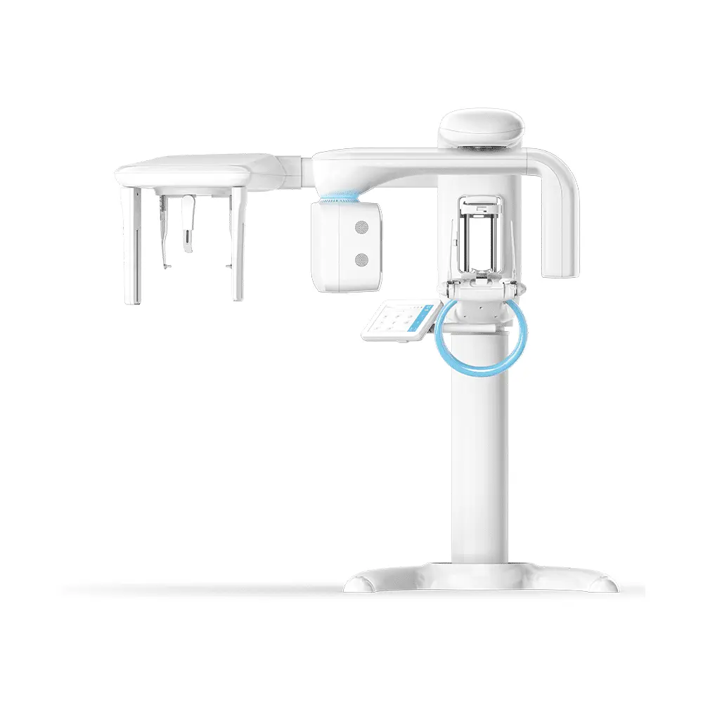
VistaVox S Ceph
3D, 2D and Ceph X-ray images with exceptional image quality
The USP of the VistaVox S Ceph is found in the ideal 3D imaging volume which is oriented to the human anatomy. The jaw-shaped field of view of the VistaVox S Ceph maps the diagnostically relevant range of a 130-mm volume and is therefore visibly larger than the most commonly used volume of Ø 80 x 80 mm. The advantage: Thanks to this changed volume shape, VistaVox S Ceph also completely covers the region of the rear molars – an essential requirement for diagnostics, e.g. for an impacted wisdom tooth. In addition to that, VistaVox S Ceph offers ten further Ø 50 x 50 mm volumes: five each for the upper jaw and for the lower jaws. These are used if the indication only requires imaging of a certain region of the jaw, e.g. for endodontical or implantological treatments. Depending on the required level of detail in the image, the volumes can be used with a resolution of either 80 or 120 μm. Supplemented by the 17 panoramic programmes in the tried-and-tested S-pan technology, this provides dental practices with excellent imaging diagnostics in both the 2D and 3D areas.
Key features:
- Ideal 3D imaging volume matched to the jaw arch (Ø 130 x 85 mm)
- Ø 50 x 50 mm volumes in 80 or 120 μm resolution
- Excellent image quality in 2D and 3D thanks to the high-resolution CsI sensor with a pixel size of 49.5 μm
- Reduced radiation dose thanks to the anatomically adapted volume
- VistaSoft – modern, ergonomic image processing software
Description
2D images with exceptional image quality
VistaVox S Ceph offers not only excellent value for money, but will also help you and your surgery team to increase your flexibility. In addition to CBCT images, the S-pan technology of the VistaVox S Ceph can be used to generate brilliant panoramic images, which set new standards in the image sharpness of extraoral images.
Key features:
- S-Pan technology for easier diagnostics
- CsI sensor for improved image quality and reduced radiation exposure
- Extremely fast, panoramic images in 7 sec.
- Tolerant of typical positioning errors – thanks to the S-Pan technology
Time-saving Cephalometric exposure with a low X-ray dose
Short scan time and high image quality with a low X-ray dose
The very short scan time of just 1.9 seconds helps to avoid motion artefacts and to reduce the radiation dose. The modern high-sensitivity CsI sensors enable excellent image quality.
3-in-1 X-ray system
In addition to the various CBCT volumes and the 17 panoramic programs, VistaVox S Ceph also offers six modes for all types of cephalometric exposures:
- Head Lateral
- Head Full Lateral
- Head PA
- SMV (submentovertex)
- Waters View
- Hand
Saving you time and money
VistaVox S Ceph is equipped with two high-end CsI sensors. The advantage: there is no need for the cumbersome process of unplugging and reconnecting between the 3D X-ray unit and the Ceph boom. To start a Ceph X-ray image acquisition, all you need to do is select the corresponding program mode.
Voltage, current60–99 kV, 4–16 mATube
Focal point0.5 mm (IEC60336)Total filtration2.8 mm ALImage detector
TypeCsI CMOS Photodiode arrayPixel size49.5 μm / 100 µmActive sensor surface135.8 x 36.4 mm / 157.2 x 16.3 mmScan times
Scan timesFrom 2 to 18 seconds for lateral head imagesCeph programmesin quick scan mode: 1.9 seconds (line scan)3D volumeØ 130 x 70 mm diagnostic
Ø 130 x 85 mm diagnostic
Ø 50 x 50 mmUnit dimensions
Height1,406–2,206 mmWeight202 kgHeight adjustment range800 mmWidth x depth1,540 x 1,190 mmInstallationWall mountingElectrical connections
Mains voltage200–240 V ACFrequency50/60 HzRated power2.2 kVA